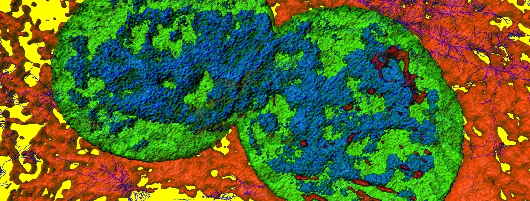
Skin - Fibroblasts
Fibroblasts are essential cellular components of the skin, that regulate and maintain the production of fibrous proteins and ground substance. These make up the extra cellular matrix (ECM), which in turn is a part of the connective tissue surrounding the cells in body tissues or organs.
Fibrous proteins
These proteins are mainly fibrin, fibronectin, elastin and collagen. They provide the basic framework or mesh required for cells to stick together, forming tissues. Collagen also provides the mechanical strength needed for tissues to hold themselves together.
Fibronectin is secreted from fibroblasts and transforms into an insoluble mesh-work that helps 'glue' the connective tissue to surrounding cells and tissues.
Collagen is considered to be the most abundant protein in the human body. It makes up 30% of all body proteins and 75% of the skin’s dry weight. It forms a major part of tissues like tendons and ligaments. The roots of the word “collagen” go back to the Greek word kólla, which means glue. True to it’s name, collagen plays a significant role in adhering cells and tissues together.
In the skin, collagen molecules in the dermis act as supporting structures that anchor cells to each other and give the skin its strength and elasticity. With age, the body begins to produce significantly less collagen, weakening the structural integrity of the skin. This results in wrinkle formation. Exposure to the UV rays in sunlight or smoking also diminishes quality collagen production, which tend to bring about signs of aging in the face, a lot faster.
Elastin is a stretchy and resilient protein. Like a rubber band, elastin helps tissues return to their previous shape after they've been stretched. In sun-damaged skin, elastin fibres become weak , which is why the skin begins to droop and sag.
Ground Substance
Ground substance contains varying amounts of water and can be either soft or firm. It contains proteins called proteoglycans which are responsible for secretion of growth factors and enzymes.
Functions of Fibroblasts during wound healing
Fibroblasts are a diverse group of cells . They have a variety of functions, within the skin itself. Cells in the superficial area of the dermis helps in the formation of the hair follicle and re-establishes the continuity of the skin during wound healing in a process called re-epithelialization. Fibroblasts in the deeper part of the skin layers, generate extracellular matrix.
Fibroblasts are activated to migrate towards a wound site to help with the healing process, where they begin to proliferate. These new fibroblasts synthesise and secrete collagen, elastin and proteoglycans which are essential for the formation of granulation tissue, to help with the healing process.
While fibroblasts are required in large numbers for initial stages of wound healing, they usually begin to disappear and are re-absorbed by the body in a process called apoptosis, during the later stages. However if this programmed cell death is delayed, it leads to excessive deposition of connective tissue and fibroblasts. This is called excessive healing or the build-up of thick scar tissue, causing the loss of normal structure and function in the affected body part. It is commonly seen as fibrosis, adhesions, keloids or hypertrophic scars.
Keloids are smooth, hard growths that occur over an injury or a scar on the skin. They can be much larger than the original wound and are commonly found on the chest, shoulders, earlobes and cheeks. They take weeks or months to form and are characterised by raised, lumpy areas of skin that itches. While they may not necessarily be harmful, keloids do raise cosmetic concerns, especially if they occur in easily visible areas.
On the other hand, when the body’s response to injury is insufficient , with few fibroblasts present and decreased generation of the connective tissue matrix, the new granulation tissue or scar tissue formed is too weak to withstand even minimal tension or pressure and begin to fall apart. This is what happens in cases of chronic, non-healing ulcers, commonly seen in people with diabetes.
References
1.Dick MK, Miao JH, Limaiem F. Histology, Fibroblast. In: StatPearls. Treasure Island (FL): StatPearls Publishing; 2021
2.Schultz GS, Chin GA, Moldawer L, et al. Principles of Wound Healing. In: Fitridge R, Thompson M, editors. Mechanisms of Vascular Disease: A Reference Book for Vascular Specialists [Internet]. Adelaide (AU): University of Adelaide Press; 2011. 23. Available from: https://www.ncbi.nlm.nih.gov/books/NBK534261
3.Varani J, Dame MK, Rittie L, et al. Decreased collagen production in chronologically aged skin: roles of age-dependent alteration in fibroblast function and defective mechanical stimulation. Am J Pathol. 2006;168(6):1861-1868. doi:10.2353/ajpath.2006.051302

